The diamond voltage imaging microscope
A new way to image voltages, that manipulates the properties of atom scale defects at the diamond surface.
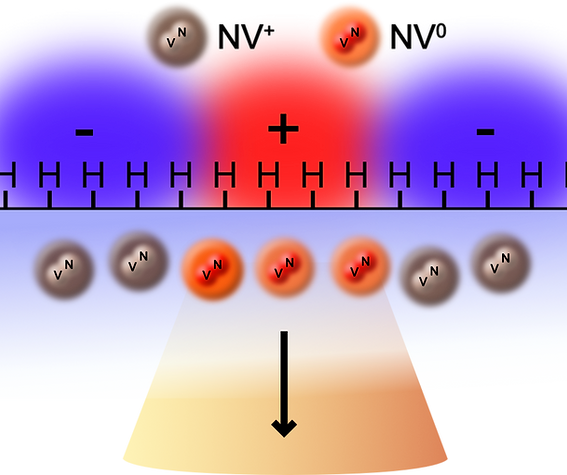
Light emitting -fluorescent- reporters in the diamond respond to the local voltage. These reporters are defects in the diamond and the color of light they emit depends what they are made of and how many electrons they have captured. The reporter on the is the NV defect, made up of one nitrogen atom and one atom sized hole (vacancy) in the diamond. When it is positively charged (missing an electron) it is dark, when it is neutrally charged it can emit orange light. By engineering the diamond surface we make it so that the charge of the reporter responds to voltages outside the diamond - transducing local voltage changes into an image.
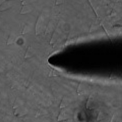
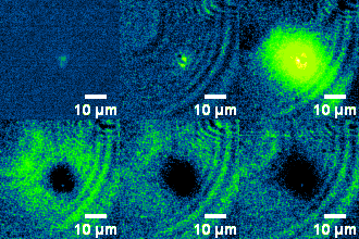
The diamond responds to change injection from an electrode. The image on the left shows an optical image of a platinum micro-electrode (2 microns in diameter at the tip). The voltage on the electrode is raised from 0V to 700mV causing an inrush of charge into the solution. As can be seen by the diamond fluorescence video on the right, the charge quickly diffuses outwards into solution. This is similar to how a cell injects ions during an action potential.